Many people have seen microscopes, but there is very little chance to see this microworld. The micrographs below are measured in micrometers, 1 micron is equivalent to 1 millionth of a meter, and the diameter of a hairline is It is about 100 microns.
Squid sucker
Squid sucker
Scanning electron micrograph of Squid Suckers. This photo was awarded the honorary prize of the 2008 International Science and Engineering Visual Contest. Loligo pealei squid has 8 arms and two tentacles, and both tentacles are covered with suction cups. The suction cup looks very much like an alien creature with a big mouth and sharp teeth. The squid straw has a diameter of about 400 microns, which ultimately allows the squid, half a metre long, to firmly control its surroundings.
Recommended reading: Plant World under the microscope (Photos)
Microcosm Photo Contest: Chicken Embryos Like Aliens (Photo)
Surprise in the micro world: the stadium can be set up on the hair of the stadium only 20 microns
2. Nano Obama
Nano Obama
The Department of Mechanical Engineering at the University of Michigan uses an electron microscope to take pictures of Obama, each made up of approximately 150 million miniature carbon nanotubes. According to John Hart of the University of Michigan, the photo was made according to the original image of Shepard Fairey, which is just over 500 microns in diameter and is made of 150 million micro-carbon nanotubes, roughly equivalent to November. The total number of US voters who voted on the day of the election on the 4th.
Recommended reading: Plant World under the microscope (Photos)
Microcosm Photo Contest: Chicken Embryos Like Aliens (Photo)
Surprise in the micro world: the stadium can be set up on the hair of the stadium only 20 microns
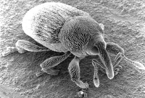
3. Weevil
Weevil
The close-up photo of the weevil photographed by the researchers using an electron microscope. The snout of the weevil is more than 100 microns in diameter.
Recommended reading: Plant World under the microscope (Photos)
Microcosm Photo Contest: Chicken Embryos Like Aliens (Photo)
Surprise in the micro world: the stadium can be set up on the hair of the stadium only 20 microns
4. Black walnut leaves
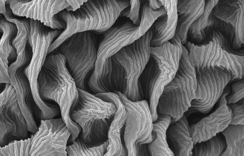
Black walnut leaves
Scanning electron micrograph of black walnut leaves. The photograph shows the cross section of the cut leaf and the upper epidermis, the mesophyll layer with the palisade cells and the vascular bundle, and the lower epidermis. The central protrusion is actually only 50 microns high.
Recommended reading: Plant World under the microscope (Photos)
Microcosm Photo Contest: Chicken Embryos Like Aliens (Photo)
Surprise in the micro world: the stadium can be set up on the hair of the stadium only 20 microns
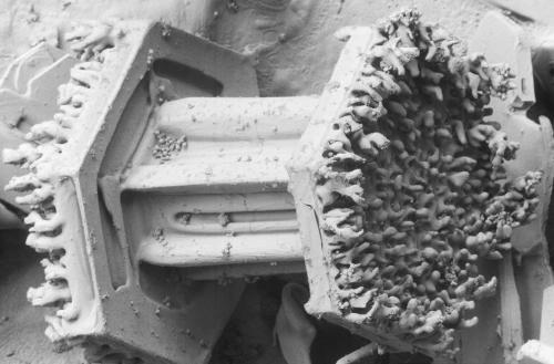
5. Transmission surface of silicon surface
Transmission gear on silicon surface
A miniature electromechanical system (MEMS) gear train made on a silicon surface with a large central gear diameter of approximately 80 microns.
Recommended reading: Plant World under the microscope (Photos)
Microcosm Photo Contest: Chicken Embryos Like Aliens (Photo)
Surprise in the micro world: the stadium can be set up on the hair of the stadium only 20 microns
6. Microalgae under the microscope
Microalgae under the microscope
Microalgae under the microscope: a plant that adsorbs carbon dioxide in the ocean.
Recommended reading: Plant World under the microscope (Photos)
Microcosm Photo Contest: Chicken Embryos Like Aliens (Photo)
Surprise in the micro world: the stadium can be set up on the hair of the stadium only 20 microns
7. Frost on columnar snow crystals
Frost on columnar snow crystals
Contact between the snow crystals and the excessively cooled water droplets in the air causes the water droplets to freeze on the crystal surface. Observations of snow crystals clearly show small droplets (about 50 microns in diameter) on the surface of the snow crystal.
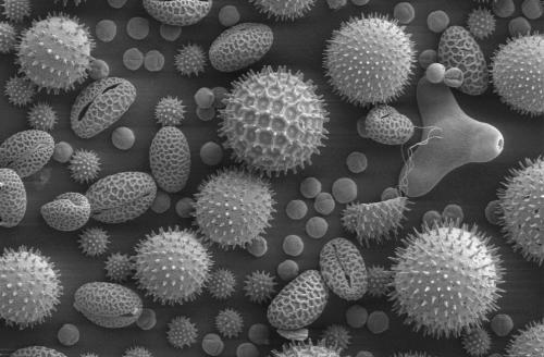
8. Various common plant pollen
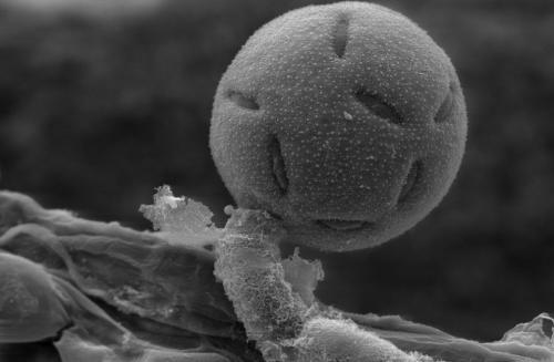
Pollen of various common plants
These include pollen from plants such as sunflowers, morning glory, hollyhocks, lilies, primroses and ramie. The largest pollen in the center is about 100 microns in diameter.
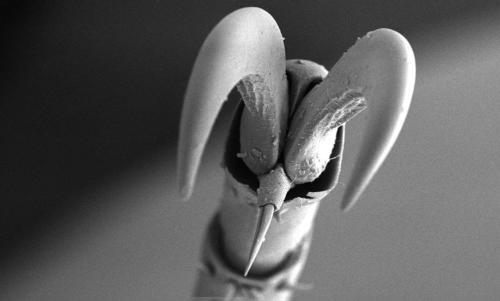
9. The legs of the fig insect
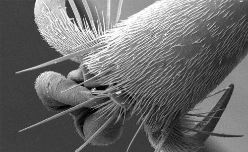
Fig insect's leg
This is a scanning electron micrograph of the tip of the paw of the adult fig insect, with a magnification of 94 times. The legs of the fig insects consist of a number of pieces, including the scorpion and the melon and conical claws seen here.
10. Mouse brain cells
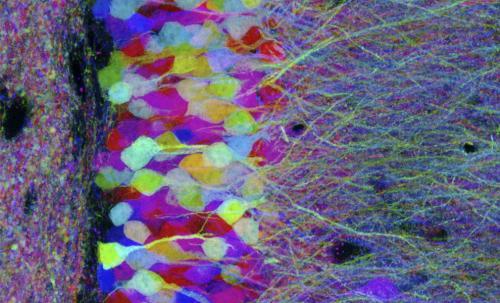
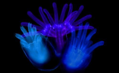
Mouse brain cells
In this photo, published on October 8, 2008, the brain cells of laboratory mice gleamed under the backdrop of colorful fluorescent proteins. Two American scientists and one Japanese scientist won the 2008 Nobel Prize in Chemistry for their work on green fluorescent protein. This breakthrough has revolutionized scientists' ability to study the normal development of diseases and living organisms.
11. The outer skin of the male scorpion
Male scorpion's chimeric bone outer skin
This electron micrograph depicts the outer skin of the crustaceous skeleton of the male scorpion with a magnification of 598 times. From this particular perspective, the exoskeleton looks like it consists of a dark gusset.
12. Ant micrograph
Ant micrograph
Scanning electron micrograph of an ant. The ant's eyes are approximately 300 microns in diameter.
13. Night moth compound eyes
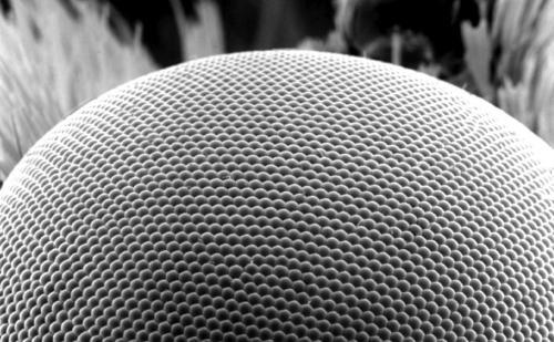
Night moth compound eye
Scanning electron micrograph of the compound eye of the night moth. Each face of the eye (ie, the small eye) is approximately 25 microns in diameter.
14. Three-dimensional photo of melanoma cells
Three-dimensional photo of melanoma cells
A three-dimensional photograph of melanoma cells, which was awarded the honorary prize of the 2008 International Science and Engineering Visual Contest. This photo was created by ion abrasion scanning electron microscopy combined with mastered data. The ion wear scanning electron microscopy method is a novel method of photographing mammalian cells at nano definition.
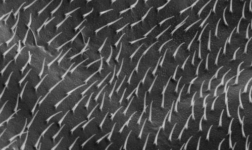
15. Arabidopsis leaves
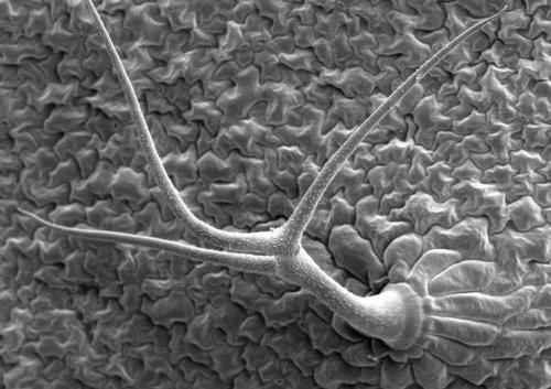
Arabidopsis leaves
Scanning electron micrographs of the lower surface of leaves of Arabidopsis thaliana showing photographs of trichomes or "leaf hairs" that grow from epidermal cells.
16. Moth head
Moth head
Scanning electron micrograph of the moth and its head side and curly nose. Its eyes are approximately 800 microns in diameter.
17. Crimson clover
Crimson clover
This scanning electron micrograph shows the ultra-fine detail seen on the surface of the "dark red clover" (the clover leaf) with a magnification of 1438.
18. Breathing holes of Drosophila larvae
Drosophila of Drosophila larvae
A scanning electron micrograph of the stomata (breathing holes) in front of the Drosophila larvae at a magnification of 1500 times.
19. Insect compound eyes
Insect compound eye
The scanning electron micrograph has a magnification of 5653 times, showing the surface of the compound eye of the unknown insect, the exposed photoreceptor cells, the supporting cells, the pigment cells, and the pigment cells constitute the hexagonal unit of the compound eye (small eye) ).
20. Hornet's legs
Bumblebee legs
This scanning electron micrograph shows the shape of the exoskeleton found on the legs of a bumblebee with a magnification of 87 times. This bumblebee has a total of six legs and was discovered on the outskirts of Decatur, Georgia. The photo shows an anatomical structure called the tarsal chain of the legs. The tibial chain is composed of a tibia, a claw, and the like.
21. Fig insect upper jaw
Fig insect maxilla
This scanning electron micrograph shows the morphological details of the tip of the maxillary protrusion of the adult fig, with a magnification of 765 times. It is precisely because of the shape of the maxillary protrusion of the fig insect that it means "helmet" in Latin. The maxillary projection is located in the center of the other, more prominent maxillary appendage. Please pay attention to the concave shape of the tip of the protrusion, as well as the many pointed protrusions in this concave surface. In nature, this protrusion is very likely to have a feeling.
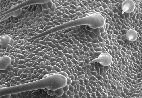
22. Amaranthus leaves
Arborvitae leaves
Scanning electron micrographs of the surface of the leaves under the genus Amaranthus show fragrant tufts and some stomata. The larger scented tufts at the bottom are approximately 50 microns in diameter.
23. Malaria mosquito tentacles
Malaria mosquito tentacles
This scanning electron micrograph shows the characteristics of the mosquito tentacles, with a magnification of 1504 times. In this unique picture, we can only see the first two fragments of the left side of the mosquito. The chitinous extension of the tentacles (called bristles) is covered with sensuous "hairs" (actually not hair), which provide feedback to the malaria mosquitoes in the presence of chemical, thermal and tactile changes.
24. Glittering mouse brain cells
Glittering mouse brain cells
In this scanning electron micrograph published by Harvard University on October 8, 2008, the brain cells of laboratory mice shimmered under the backdrop of colorful fluorescent proteins. Two American scientists and one Japanese scientist won the 2008 Nobel Prize in Chemistry for their work on green fluorescent protein. This breakthrough has revolutionized scientists' research into the normal development of diseases and living organisms.
25. Beetle head
Beetle head
This scanning electron micrograph shows some exoskeleton morphological features of the head of an unknown beetle with a magnification of 58. The hair we see is actually a sensory organ called “bristle†that provides the beetle with all the information about environmental changes, such as temperature, wind direction, chemical alignment, and so on.
26. Mosquito tentacles
Mosquito tentacles
This scanning electron micrograph shows the first fragment of the "scape" or the left side of the mosquito, with a magnification of 500. Note that at the center of the whisker is a concave surface, and the second segment of the shank, where the stem is connected to the other segments. The small eye of the grape shape around the handle is the functional unit of the compound eye.
27. Star flower pollen
Star flower pollen
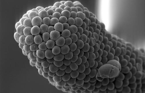
Scanning electron micrograph of pollen grains of Penta lanceolata flower. The pollen grains have a diameter of about 40 microns.
28. Star flower head
Star flower head
Scanning electron micrograph of the stigma of the star, with a stigma diameter of approximately 140 microns.
29. Schistosomiasis
Schistosomiasis
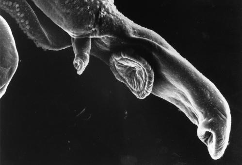
This is a scanning electron micrograph of the parasite schistosomiasis. After a person comes into contact with water infected with schistosomiasis, the schistosomiasis enters the human body through the skin of the person. Adult schistosomiasis usually parasitizes in the blood vessels of the host. This photo has a magnification of 256 times.
30. Breast cancer cells
Breast cancer cell
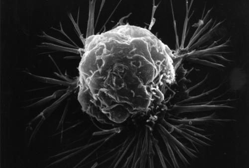
Scanning electron micrograph of breast cancer cells. This photo shows the overall shape of the cancer cell surface with very high magnification. Cancer cells are so clearly presented to us through internal details, but studies using scanning electron microscopy can show how cells respond to changing environments while showing the distribution of hormones and other biomolecule binding sites.
Beijing Khan Meng Zixing Instrument Co., Ltd. ()
Tuna Loin
Tuna Loin,Frozen Tuna Loin,Fresh Tuna Loin,Yellowfin Tuna Loin
ZHEJIANG RETRONX FOODSTUFF INDUSTRY CO.,LTD , https://www.retronxfoods.com